
Gallbladder Diseases
Functions of Gallbladder
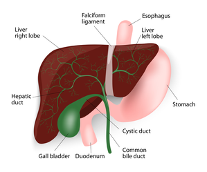
The gall bladder is a small pear-shaped organ attached to the side of the bile duct by a small secondary duct, the cystic duct. The bile duct is a tube that carries bile from the liver to the small intestine. When the patient is fasting, the lower end of the bile duct closes and bile back-flows into the gall bladder. There it is concentrated by the gall bladder, absorbing the water in the bile. When a patient eats a fatty meal, the gall bladder squeezes out the bile to help absorb the fats. If stones are present, they can cause the gall bladder to go into spasm and causes severe pain as well as nausea, vomiting, bloating, or fever. If a gallstone travels down the bile duct, you could have liver dysfunction, bile duct infection or inflammation in the pancreas, called pancreatitis. The only way to prevent this and the other problems that can occur with gall stones is to remove the gall bladder. Since the gall bladder is only one of the mechanisms of fat digestion, its removal does not cause any major interference with the patient’s digestive process. In many cases of patients with stones, the gall bladder is not functioning and so digestion of fats is not affected by its removal.
Gallstones
If the bile contains too much cholesterol or bilirubin, or the gallbladder does not empty properly, then stones can form within the gall bladder. Gallstones can be cholesterol stones, pigment stones or a mixture of the two. Cholesterol stones are largely made of solidified cholesterol. Pigment stones are dark stones made of bilirubin. The majority of stones are mixed stones that contain cholesterol as well as pigment. The gallbladder can develop just one or two large stones (some can be as large as a golf ball), or lots of tiny stones (as small as grains of sand).
Causes of Gallstones
The gallbladder is a small organ in the right upper abdomen under the liver. When healthy, it stores some of the bile that is made by the liver. Bile is necessary to digest and absorb the fats in the foods we eat. When there is an imbalance of the chemicals in the bile, gallstones can form.
A number of causes have been suggested. It is thought that some people secrete more cholesterol than others. As the gallbladder concentrates the bile stored in it, the cholesterol precipitates forming crystals and that these adhere together forming the stones. The stones then tend to enlarge or multiply especially if there is any infection involved. Pregnancy, obesity, weight loss and a family history of gallstones are factors that increase the chances of developing gallstones.
Risk Factors for Gallstones
Women, particularly between the ages of 20 and 60 years, are more likely to form gallstones than men. In general, those over 60 (men and women) are at a higher risk of developing gallstones. People who are overweight are more likely to form gallstones. Excess estrogen from numerous pregnancies, hormone replacement therapy, or birth control pills may increase cholesterol levels in bile, slow down gallbladder emptying, and lead to gallstones.
People who have biliary infections (for example liver flukes in the tropics) can develop gallstones. Individuals with hereditary blood disorders such as sickle cell anemia (in which too much bilirubin is formed due to the breakdown of blood cells) are more likely to form pigment stones. Going on a diet (with rapid weight loss) and certain cholesterol-reducing drugs can also increase the risk of gallstone formation. A high level of cholesterol in the blood is not necessarily a factor in the development of gallstones. Many people have a “family history” of gallstones but there is no particular gene that has been associated with this.
Prevention for Gallstones
There is no special diet you can follow, particular foods you should avoid, or medications you can take to specifically prevent the formation of gallstones. Those who already have pain from gallstones often find that fatty or oily food can trigger the pain. So a low-fat diet can help keep the pain at bay. Gallstones are not related to stones in other parts of the body, particularly stones in the kidneys or in the urinary bladder.
Symptoms of Gallstones
The main symptom is pain, known as gallstone or biliary colic. This commonly occurs in the mid upper abdomen or under the right ribs. It tends to radiate around the rib margin and into the back. It can be precipitated by eating fats. It is severe and can last some hours. The pain usually goes but frequently recurs. In some cases infection sets in, cholecystitis, and the patient develops severe pain under the right ribs with fever. Intravenous antibiotics are necessary to treat the infection and the problem usually takes 3-4 days to settle. If a gallstone passes down the cystic duct into the bile duct it can block the flow of bile leading to jaundice. This is a surgical emergency requiring removal of the obstruction, especially if infection sets in. A stone in the bile duct may also cause inflammation of the pancreas causing a serious condition known as pancreatitis. Rarely, if the stones are left for many years, cancer of the gall bladder may develop.
Often, gallstones remain “silent” and do not cause any symptoms at all. Silent gallstones are usually discovered by accident when tests are done for other problems. The milder symptoms of gallstones include abdominal bloating, belching, indigestion, and nausea, usually after a meal. More severe symptoms include attacks of abdominal pain and vomiting. The pain is usually in the upper abdomen, often more to the right, and can move to the right shoulder blade or shoulder tip. It may come on after meals, especially with fatty foods. This kind of pain is called biliary colic. Most attacks of biliary colic settle after a few hours.
Gallstone can cause symptoms similar to those of a heart attack, appendicitis, bowel obstruction, peptic ulcer, hiatus hernia, pancreatitis, hepatitis and occasionally biliary cancer. It is therefore very important that the correct diagnosis is made.
Diagnosis of Gallstones
To determine if you have gallstones, you will likely undergo an ultrasound, which uses sound waves to detect gallstones and evaluate the bile ducts. If there are abnormalities in the pancreas or bile ducts, additional imaging tests including a CAT scan or magnetic resonance cholangiopancreatography (MRCP), may be necessary. Blood tests can also be helpful if infection or bile duct blockage is suspected.
Complications Caused by Gallstones
Gallstones can lead to acute inflammation of the gallbladder (cholecystitis). Acute cholecystitis is a medical emergency and requires admission to hospital. Treatment has conventionally involved pain killers and antibiotics to settle the acute inflammation, followed six weeks later by an operation to remove the gall bladder. Current best practice involves operating immediately with laparoscopic surgery and this has lead to better outcomes.
Acute cholecystitis caused by aggressive bacteria can cause the gall bladder wall to rot and disintegrate, and the gall bladder may then burst into the abdominal cavity or into an adjacent bit of bowel.
If there have been repeated attacks of inflammation, the gall bladder becomes chronically inflamed, shrunken and scarred. This is called chronic cholecystitis. If the outflow from the gall bladder is totally blocked by a stone it may become a bag full of stagnant bile (a mucocoele) or a bag of pus (empyema).
Gallstones slipping out of the gallbladder into the bile duct can block the flow of bile and cause obstructive jaundice. If you have severe abdominal pain, chills, fever, yellow discoloration of the eyes or skin, or pale stools, you should urgently consult a doctor.
Gallstones passing down the bile duct may also cause inflammation of the pancreas, Acute pancreatitis is a medical emergency, which occasionally escalate into life-threatening complications.
Gallstones Vs. Gallbladder Cancer
Although gallstones are a common problem, gallbladder cancer is a rare form of cancer except in certain parts of the world and in some ethnic groups. Underlying genetic or dietary factors may be the reason for this.
Gallstones are a risk factor for gallbladder cancer, though the mechanism is unclear. Usually patients with large stones of long duration are at increased risk for cancer. More than the stones themselves, incomplete emptying of the gallbladder and the chronic inflammation that goes with it may have a role to play in initiating the cancer.
Calcium deposits in the wall of the gallbladder (a consequence of chronic inflammation), described as Porcelain gallbladder, increase the risk of gall bladder cancer. The picture on this page shows a CT scan, with a porcelain gall bladder clearly visible (you can click on the picture to enlarge it).
Chronic Salmonella infection of the gallbladder, which predisposes to gallstone formation, can predispose to gallbladder cancer.
Patients with choledochal cysts (a focal or diffuse bulge in the bile duct) or abnormalities at the point where the pancreatic and bile ducts join and enter the bowel are also known to be at increased risk for gall bladder or bile duct cancer.
Sometimes patients are found to have polyps (little growths) on the lining of the gallbladder. Polyps are generally benign, but patients with a single, large (> 1cm) polyp are more likely to develop cancer within their polyp. See below for more information about gall bladder polyps.
Gallbladder Polyps
Polyps are growths on the inner lining of the gallbladder wall. Gallbladder polyps are often discovered incidentally on scans, and not all of them warrant removal. Polyps that are single, sessile (do not have a stalk), and are 1 cm or more in diameter should be deemed as suspicious i.e. they may develop into cancerous (malignant) growths. Also, polyps that have developed in older patients (over the age of fifty years), developed in association with stones or are associated with symptoms, are at a higher risk of being malignant or subsequently turning malignant. Laparoscopic cholecystectomy (keyhole surgery to remove the gall bladder) is the treatment of choice in these patients, unless the suspicion of malignancy is high, in which case open exploration is preferable, with preparation for extended resection if necessary.
For polyps that are deemed low-risk, Mr Karametos's practice is to recommend follow-up at first with regular ultrasound scans every 6-12 months, which can stop after 2 years if they remain unchanged.
Other Problems With Similar Symptoms
The symptoms of gallstones can be similar to those of heart disease, appendicitis, bowel obstruction, peptic ulcer, hiatus hernia, pancreatitis, hepatitis and occasionally biliary cancer. It is therefore very important that the correct diagnosis is made. Other tests may be required, including an Gastroscopy to look inside the oesophagus and the stomach.
Read More About...